A groundbreaking study using PET scans has revealed that autistic individuals have fewer brain synapses, directly correlating with more pronounced autism traits like social and communication challenges.
This discovery, illustrating clear links between brain structure and behavioral expressions, could revolutionize diagnostic approaches and enhance support mechanisms, potentially leading to more targeted interventions and improved quality of life for those on the autism spectrum.
Synaptic Research in Autism
For decades, researchers have studied animal models and post-mortem brain tissue to explore social and communication differences that define autism. Now, a groundbreaking study has identified a molecular difference in the brains of living autistic people, linked to core features of the condition. This is the first time synaptic density has been measured in living individuals with autism.
Breakthrough in Live Brain Analysis
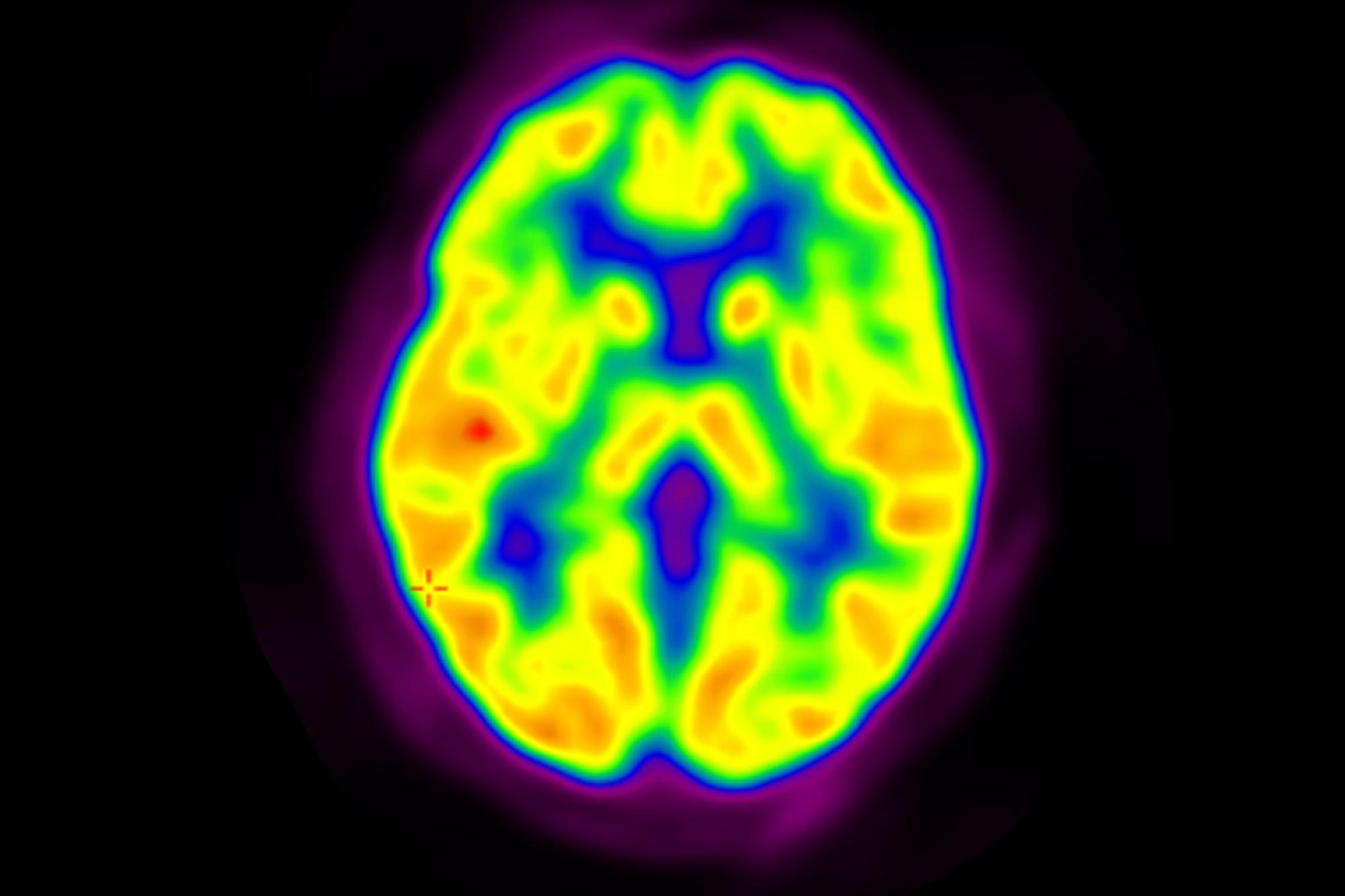
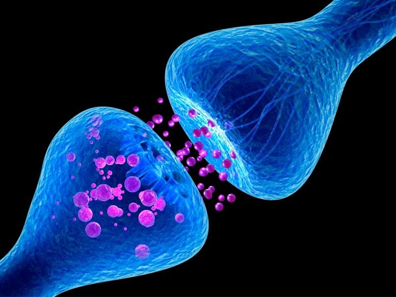
Using positron emission tomography (PET) scans, researchers discovered that autistic adults have fewer synapses—essential junctions where nerve cells transmit signals—compared to neurotypical individuals. They also observed that lower synapse counts correlated with more pronounced autistic traits. The team recently published their findings in Molecular Psychiatry.
“As simple as our findings sound, this is something that has eluded our field for the past 80 years,” says James McPartland, PhD, Harris Professor in the Yale Child Study Center and the study’s principal investigator. “And this is truly remarkable—because it’s very unusual to see correlations between brain differences and behavior this strong in a condition as complex and heterogenous as autism.”
There are several different theories about brain differences in autistic individuals, and atypical connectivity has been at the root of a number of these hypotheses. This has made synapses a prime area to investigate. “Synapses are the way neurons communicate,” says Adam Naples, PhD, assistant professor in the Child Study Center and co-investigator on the study. “They’re the fundamental mechanism for how information moves around the brain and is computed.”
Previous studies have measured synaptic connectivity in indirect ways such as through animal models or post-mortem studies. “It’s like trying to figure out what something is by looking at the shadow it casts on the wall,” says McPartland. But the introduction of a new element in the PET scanning protocol permitted McPartland and his team to see connectivity directly—in living human beings—for the very first time.
PET Scans Reveal Fewer Synapses in Autistic Brains
Before the study, all of the subjects participated in a clinician interview. Given the complexity of autism, clinicians used the Autism Diagnostic Observation Schedule (ADOS)—the gold standard for diagnosing autism—to evaluate participants for the condition. Participants also filled out self-report questionnaires about their own experiences living with autism, such as difficulty with social interactions or sensory issues. The researchers ruled out any potential subjects with medical conditions or neuropsychiatric disabilities that could influence the study’s findings. In total, 12 autistic adults and 20 neurotypical adults participated in the research.
“As simple as our findings sound, this is something that has eluded our field for the past 80 years.”
James McPartland, PhD
Then, each participant underwent a brain scan using both magnetic resonance imaging (MRI) and PET technology. The MRI scan allowed researchers to visualize each participant’s brain anatomy in great detail. Before the PET scan, researchers injected a novel radiotracer known as 11C‑UCB‑J, which was developed with the Yale PET Center, and which enabled them to measure synaptic density in the brain.
The researchers found that autistic people had 17% lower synaptic density across the whole brain compared to neurotypical individuals. Furthermore, they found that lower synaptic density was significantly correlated to the number of social-communication differences, such as reduced eye contact, repetitive behaviors, and difficulty understanding social cues, in these individuals. In other words, the fewer synapses a person had, the greater number of autistic traits they showed.
New Insights Into Autism Diagnosis and Treatment
A major limiting factor in clinicians’ ability to understand and offer support for autistic people, says McPartland, is the lack of a mechanistic understanding of the condition. “Today’s diagnostic criteria [which predate this new study] involve descriptions of behavior that are broad and pretty vague,” he explains. “We could be so much more effective in figuring out whether and what supports are needed if we could aid our clinical decisions with an understanding of the biology of autism.”
Investigating the underlying mechanisms of autism could also help researchers better define subgroups within the condition. “We historically had the hubris to think we could create subgroups without this understanding,” McPartland says. The Diagnostic and Statistical Manual of Mental Disorders, 4th edition (DSM-IV), which was published in 1994, divided autistic spectrum disorders (ASD) into Asperger’s syndrome, pervasive developmental disorder not otherwise specified (PDD-NOS), and autistic disorder. “Then in the DSM-5, we had to swallow our pride and throw these categories away because they weren’t working.”
Today, the DSM-5 defaults to one broad, non-specific category of ASD. McPartland hopes his work will help pave the way for parsing autism into better-defined subgroups, which will in turn help clinicians better understand the wide range of features autistic individuals may present with.
It is still unclear whether autistic people are born with fewer synapses, or if this difference occurs as a result of living with autism. But PET scans could one day potentially help clinicians anticipate a child’s prognosis and enable the care team to administer appropriate interventions earlier. “This is the dream — to be able to give biologic confirmation to patients and their families,” says David Matuskey, MD, associate professor of radiology & biomedical imaging, and the study’s first author. “That would change everything.”
Advancements and Goals for Future Autism Research
In future studies, the team is investigating the use of nonradioactive approaches that are less expensive than PET scans for directly studying the autistic brain. They are also interested in measuring synapses in adolescent brains to better understand how this may evolve as an individual ages. Finally, the team plans to explore how their findings relate to other outcomes associated with autism. For instance, autistic individuals are at a higher risk of mental health issues such as depression or anxiety than neurotypical people. “This is something that’s really important for us to investigate to serve our overarching goal, which is to get information that can maximize the quality of life for autistic people,” says McPartland.
Reference: “11C-UCB-J PET imaging is consistent with lower synaptic density in autistic adults” by David Matuskey, Yanghong Yang, Mika Naganawa, Sheida Koohsari, Takuya Toyonaga, Paul Gravel, Brian Pittman, Kristen Torres, Lauren Pisani, Caroline Finn, Sophie Cramer-Benjamin, Nicole Herman, Lindsey H. Rosenthal, Cassandra J. Franke, Bridget M. Walicki, Irina Esterlis, Patrick Skosnik, Rajiv Radhakrishnan, Julie M. Wolf, Nabeel Nabulsi, Jim Ropchan, Yiyun Huang, Richard E. Carson, Adam J. Naples and James C. McPartland, 4 October 2024, Molecular Psychiatry.
DOI: 10.1038/s41380-024-02776-2
