
The membrane that surrounds cells in living organisms is extremely flexible and sensitive. How it protects itself from damage and renews itself is crucial for many life processes, and is not yet fully understood in detail. Scientists at Forschungszentrum Jülich have now been able to gain fascinating new insights using cryo-electron microscopy.
Their work is published in the journal Nature Structural & Molecular Biology.
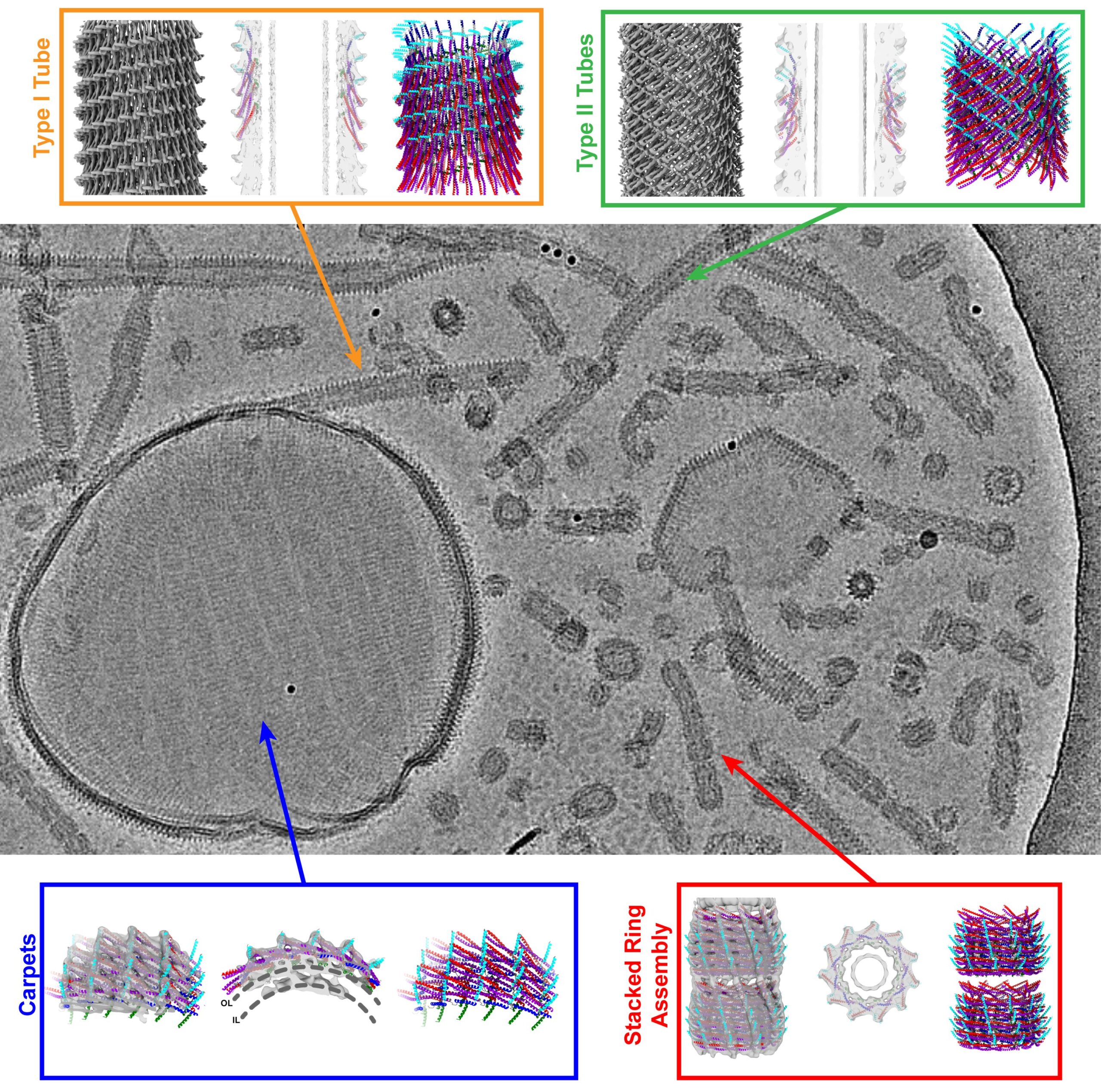
The membrane protein Vipp1, known from the photosynthetic apparatus of plants, algae, and bacteria, can form various structures that could serve as tools to stabilize the cell membrane and strengthen it if necessary.
In a second study published in the same journal, the researchers were also able to gain new insights into the function of the related protein PspA, which is found in bacteria. Both molecules, Vipp1 and PspA, are unusually plastic and can adopt different structures, creating rings and tubes with different diameters.
The cell membrane has numerous important functions. For instance, it protects the inside of the cell from the environment. At the same time, nutrients are absorbed through the cell membrane, waste products are excreted, and signals are transmitted between cells.
Despite its central role, the cell membrane is also very sensitive. It consists of a thin layer of lipids that—although protective by themselves—are also susceptible to stress caused by physical pressure and stretching or chemical influences. Environmental factors such as UV radiation or toxins can also damage the membrane.
In plant cells, for example, intense light can severely stress and even damage the membranes in the chloroplasts, where photosynthesis takes place. Proteins such as Vipp1 are therefore essential for the survival of the cell, as they protect the membrane structures and repair them if necessary.
How exactly the mechanism works is not yet fully understood. However, thanks to the state-of-the-art cryo-electron microscopes, the researchers have now been able to gain new insights into the interaction between Vipp1 and the cell membrane. They discovered that Vipp1 forms carpet-like structures on the cell membrane and stabilizes it. In addition, they found ring complexes and tubes made of Vipp1 filled with membrane, which can possibly “pinch off” damaged membrane areas as well as connect two separate membranes.
These findings provide new insights into the ability of the proteins Vipp1 and PspA to alter cell membranes and thus protect vital processes in the cells. These discoveries could contribute to the development of new biotechnological applications in the future, such as the production of biomaterials or the optimization of photosynthesis in plants.
Vipp1 is particularly important, as it is involved in the formation and maintenance of thylakoid membranes—membranes in the chloroplasts of plant cells where the light reaction of photosynthesis takes place, i.e., the conversion of light into chemical energy.
It is interesting to note that the basic mechanism is highly similar to the ESCRT-III proteins, which are also highly conserved in human cells. These proteins have remained essentially unchanged in the course of evolution, which indicates an important function. A better understanding of the structure and function of these proteins could thus lead to the development of new drugs, such as antibiotics, that target the processes in cellular membranes.
Cryo-electron microscopes from Forschungszentrum Jülich’s Ernst Ruska-Centre (ER-C) were used in both studies. The microscopes enabled the researchers to study the proteins in atomic resolution and to observe them in an unusually high number of structural states as well as the interactions between the proteins and the membranes. These studies are part of an established collaboration with the research group of Prof. Dr. Dirk Schneider from the Johannes Gutenberg-University Mainz.
More information:
Benedikt Junglas et al, Structural basis for Vipp1 membrane binding: from loose coats and carpets to ring and rod assemblies, Nature Structural & Molecular Biology (2024). DOI: 10.1038/s41594-024-01399-z
Benedikt Junglas et al, Structural plasticity of bacterial ESCRT-III protein PspA in higher-order assemblies, Nature Structural & Molecular Biology (2024). DOI: 10.1038/s41594-024-01359-7
Provided by
Forschungszentrum Juelich
Citation:
Cryo-electron microscopy provides new insights into the cell’s repair system (2024, October 8)
retrieved 8 October 2024
from https://phys.org/news/2024-10-cryo-electron-microscopy-insights-cell.html
This document is subject to copyright. Apart from any fair dealing for the purpose of private study or research, no
part may be reproduced without the written permission. The content is provided for information purposes only.