
Researchers have developed a groundbreaking tool that offers new insights into the brain’s extracellular matrix (ECM), revealing how it changes over time and interacts with neurons during development, cognitive processes, and aging.
This tool uses a combination of proteins and fluorescent dyes to visualize and track the dynamic structure of ECM in rodent brain cells.
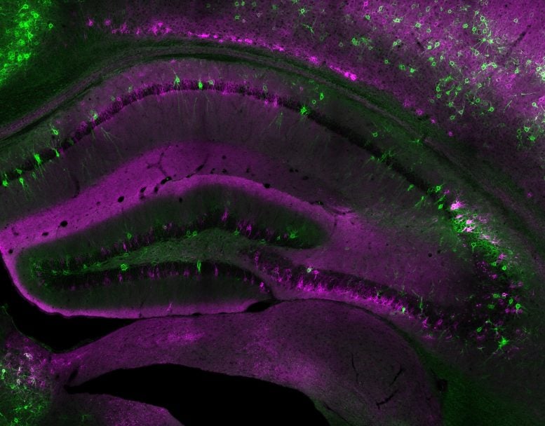
Brain Extracellular Matrix
Between and around the billions of neurons in the human brain is an equally vital scaffold, the extracellular matrix (ECM). An interlinked net of proteins and sugars that surrounds brain cells, the ECM is more than simple structural support; changes in the ECM can regulate complex brain functions including memory, learning, and behavior. However, studies of the brain’s ECM have been limited by the lack of tools to observe dynamic changes to its structure.
Now, a new genetic labeling tool developed by University of Utah Health researchers Igal Sterin, PhD, and Sungjin Park, PhD, has revealed new patterns in brain ECM in mice, including differences in the amount of matrix deposited on different types of neurons. A major advantage of the tool is that it can detect changes in the ECM over time, giving new insights into how the brain develops.
How the New Tool Works
The new tool has two main parts: a protein that binds to the main ingredients of the brain ECM, fused to another protein that irreversibly sticks to a variety of synthetic fluorescent dyes. When scientists introduced the tool into neurons, it bound to the surrounding matrix; then, they added a fluorescent dye to make matrix structures visible. Using this tool, the researchers were able to watch as ECM was deposited over time in cultured rodent brain cells, making out dense clusters of matrix that appeared on only certain neurons and at different times.
Implications for Future Brain Studies
The researchers were also able to measure changes in brain ECM in mice by adding different fluorescent dyes at different times. They could infer that ECM structures that were marked by the second dye, but not the first, had developed in the time between the two dyes were added.
The scientists hope that this tool will open the door to many further studies of how the ECM contributes to brain function in complex ways. “In the brain, the ECM regulates neuronal plasticity and cognitive function, but its structural features remain poorly understood,” Park says. “By using our longitudinal genetic tool to track ECM dynamics, we can visualize how neurons assemble and remodel ECM during development, cognitive processes, and aging.”
Reference: “Dynamic organization of neuronal extracellular matrix revealed by HaloTag-HAPLN1” by Igal Sterin, Ava Niazi, Jennifer Kim, Joosang Park and Sungjin Park, 8 September 2024, Journal of Neuroscience.
DOI: 10.1523/JNEUROSCI.0666-24.2024