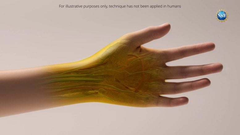
Using common food dye, researchers make skin and muscle safely and reversibly transparent.
Scientists at Stanford University have developed a groundbreaking technique using food-safe dye to make animal tissues transparent, enhancing the visibility of internal organs. This innovation has potential applications ranging from medical diagnostics to cancer treatment and has shown promising results in both theoretical and practical tests.
Groundbreaking New Imaging Technique Unveiled
Researchers have developed a new way to see organs within a body by rendering overlying tissues transparent to visible light.
The counterintuitive process—a topical application of food-safe dye—was reversible in tests with animal subjects, and may ultimately apply to a wide range of medical diagnostics, from locating injuries to monitoring digestive disorders to identifying cancers.
Stanford University researchers published the research ″Achieving optical transparency in live animals with absorbing molecules″ in the September 6, 2024, issue of Science.
″Looking forward, this technology could make veins more visible for the drawing of blood, make laser-based tattoo removal more straightforward, or assist in the early detection and treatment of cancers,″ said Stanford University assistant professor of materials science and engineering Guosong Hong, a U.S. National Science Foundation CAREER grantee who helped lead this work. ″For example, certain therapies use lasers to eliminate cancerous and precancerous cells, but are limited to areas near the skin’s surface. This technique may be able to improve that light penetration.″
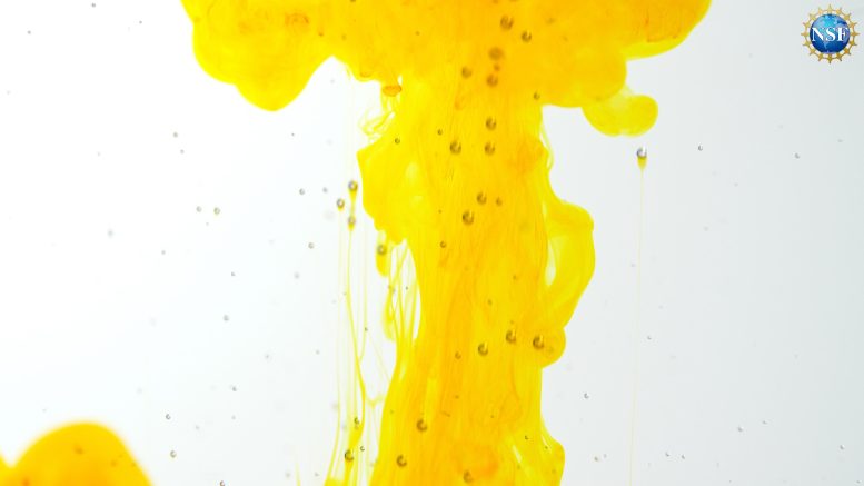
Researchers at Stanford University have developed a way to make skin and other tissues transparent using a simple food dye, a reversible technique with potential for revolutionizing internal medicine. In this clip, thin slices of chicken breast become transparent on exposure to the dye FD & C Yellow 5. Credit: U.S. National Science Foundation
An Illuminating Solution
To master the new technique, the researchers developed a way to predict how light interacts with dyed biological tissues.
Those predictions required a deep understanding of light scattering, as well as the process of refraction, where light changes speed and bends as it travels from one material into another.
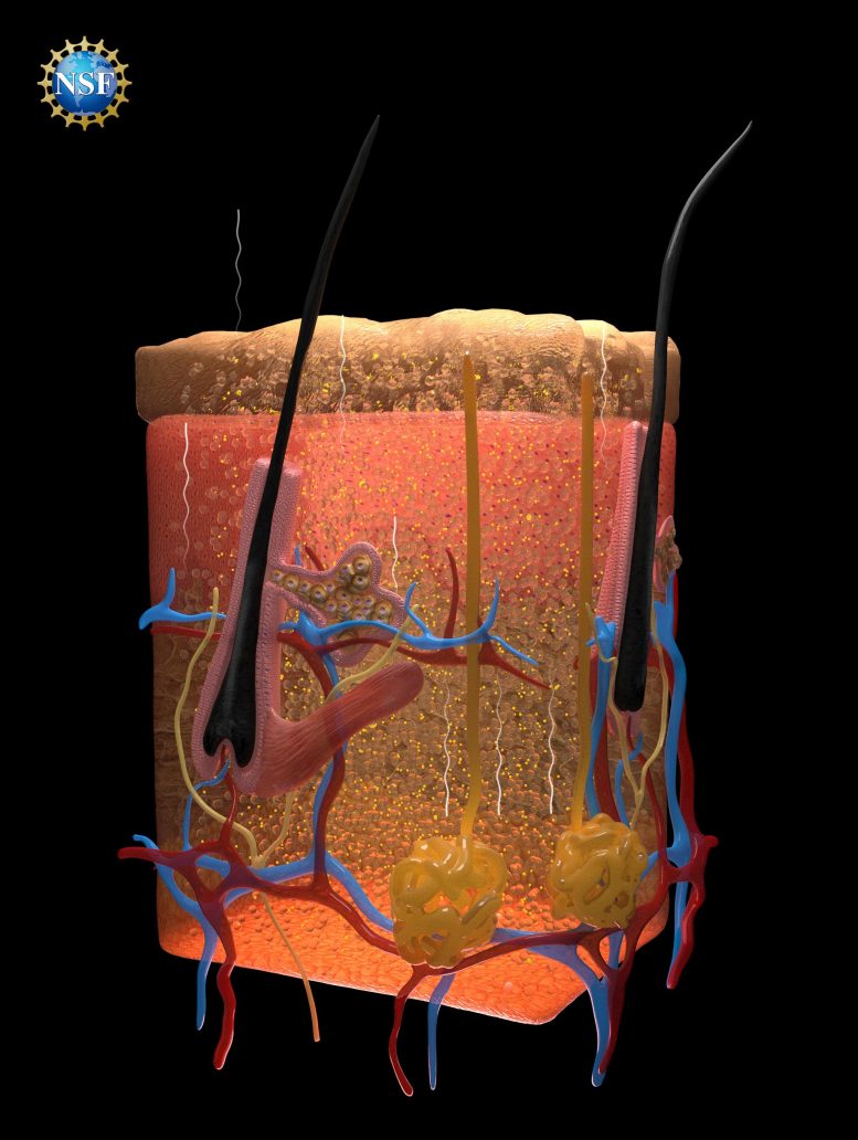
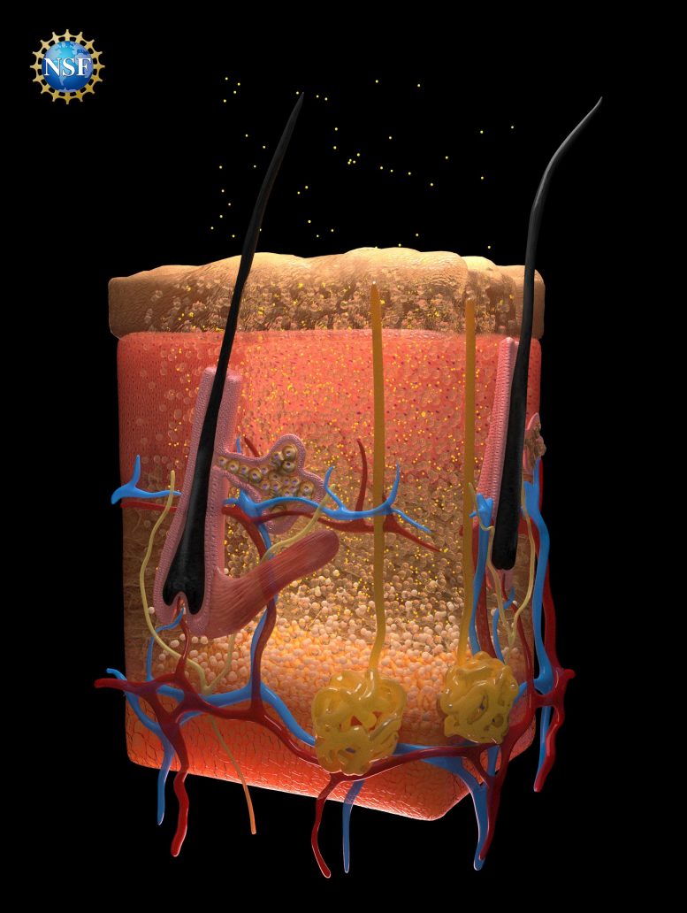
Scattering is the reason we cannot see through our body: Fats, fluids within cells, proteins, and other materials each have a different refractive index, a property that dictates how significantly an incoming light wave will bend.
In most tissues, those materials are closely compacted together, so the varied refractive indices cause light to scatter as it passes through. It is the scattering effect that our eyes interpret as opaque, colored, biological materials.
The researchers realized if they wanted to make biological material transparent, they had to find a way to match the different refractive indices so light could travel through unimpeded.
Breakthrough With Tartrazine
Building upon fundamental insights from the field of optics, the researchers realized dyes that are the most effective at absorbing light can also be highly effective at directing light uniformly through a wide range of refractive indices.
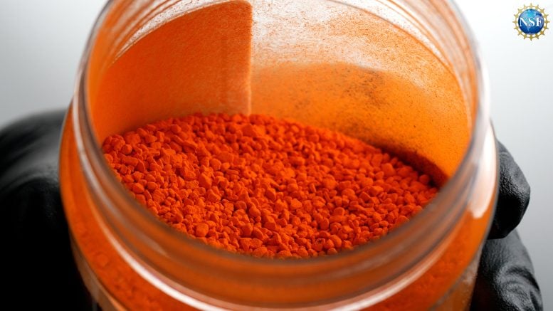
One dye the researchers predicted would be particularly effective was tartrazine, the food dye more commonly known as FD & C Yellow 5. It turns out, they were correct: When dissolved into water and absorbed into tissues, tartrazine molecules are perfectly structured to match refractive indices and prevent light from scattering, resulting in transparency.
From Theory to Practice
The researchers first tested their predictions with thin slices of chicken breast. As tartrazine concentrations increased, the refractive index of the fluid within the muscle cells rose until it matched the refractive index of the muscle proteins – the slice became transparent.
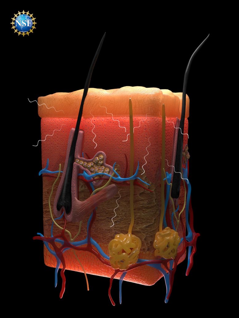
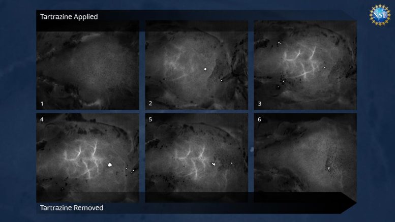
Then, the researchers gently rubbed a temporary tartrazine solution on mice. First, they applied the solution to the scalp, rendering the skin transparent to reveal blood vessels crisscrossing the brain. Next, they applied the solution to the abdomen, which faded within minutes to show contractions of the intestine and movements caused by heartbeats and breathing.
The technique resolved features at the scale of microns, and even enhanced microscope observations. When the dye was rinsed off, the tissues quickly returned to normal opacity. The tartrazine did not appear to have long-term effects, and any excess was excreted in waste within 48 hours.
The researchers suspect that injecting the dye should lead to even deeper views within organisms, with implications for both biology and medicine.
Old Formulas Yield New Window Into Medicine
Supported by a range of federal and private grants, the project began as an investigation into how microwave radiation interacts with biological tissues.
In exploring optics textbooks from the 1970s and 1980s, the researchers found two key concepts: mathematical equations called Kramers-Kronig relations and a phenomenon called Lorentz oscillation, where electrons and atoms resonate within molecules as photons pass through.
Well studied for more than a century, yet not applied to medicine in this way, the tools proved ideal for predicting how a given dye can raise the refractive index of biological fluids to perfectly match surrounding fats and proteins.
Graduate researcher Nick Rommelfanger, working under an NSF Graduate Research Fellowship, was one of the first to realize that the same modifications that make materials transparent to microwaves could be tailored to impact the visible spectrum, with potential applications in medicine.
Animation depicting the tissue transparency effect and how it might appear if tested with humans in the future. The latter part of the animation shows how photons interact with tissues at the cellular level, both with and without FD & C Yellow 5 saturation. Credit: Keyi “Onyx” Li/U.S. National Science Foundation
Harnessing Old Equipment for New Discoveries
Transitioning from theory to experimentation, postdoctoral researcher Zihao Ou—the study’s lead author—ordered a number of strong dyes and began the process of meticulously evaluating each for ideal optical properties.
Ultimately, the team grew to 21 students, collaborators, and advisors, involving several analytical systems.
One that proved critical was a decades-old ellipsometer nestled among newer equipment at the Stanford Nano Shared Facilities, part of the NSF National Nanotechnology Coordinated Infrastructure (NNCI). The ellipsometer is a tool familiar to semiconductor manufacturing, not biology. However, in a possible first for medicine, the researchers realized it was perfect to predict the optical properties of their target dyes.
″Advanced research facilities constantly aim to strike the right balance by providing access to basic tools and expertise while making space for newer, larger, and more powerful instrumentation,″ said NSF Program Officer Richard Nash, who oversees the NSF NNCI. ″While a basic workhorse such as an ellipsometer would rarely make headlines, it nevertheless can play a crucial role when deployed for atypical uses like the case here. Open access to such instrumentation is foundational for making groundbreaking discoveries, as those instruments can be deployed in new ways to generate fundamental insights about scientific phenomena.″
With methods grounded in fundamental physics, the researchers hope their approach will launch a new field of study matching dyes to biological tissues based on optical properties, potentially leading to a wide range of medical applications.
″As an optics person, I’m amazed at how they got so much from exploiting the Kramers-Konig relationship,″ said NSF Program Officer Adam Wax, who has supported Hong’s work. ″Every optics student learns about them, but this team has used the equations to figure out how a strongly absorbing dye can make skin transparent. Using an NSF EAGER grant, Hong was able to step out in a bold new direction, a great example of how fundamental optics knowledge can be used to create new technologies, including in biomedicine.″
Instrumental NSF Support
″NSF′s support played an instrumental role in the success of this work,″ added Hong. ″The NSF CAREER award was my first major funding, and it arrived at a particularly challenging time, during the darkest moments of the pandemic. My lab faced significant difficulties generating data due to the shutdown, and the award was a vital springboard, enabling me to pursue some of our most exciting and innovative projects – including the research that culminated in this Science paper. The flexibility and encouragement from the NSF awards were crucial in keeping me on track and allowed me the freedom to explore new and uncharted territories in my field.″
Please note: The technique described above has not been tested on humans. Dyes may be harmful. Always exercise caution with dyes and do not consume directly, apply to people or animals, or otherwise misuse.
For more on this research:
Reference: “Achieving optical transparency in live animals with absorbing molecules” by Zihao Ou, Yi-Shiou Duh, Nicholas J. Rommelfanger, Carl H. C. Keck, Shan Jiang, Kenneth BrinsonJr, Su Zhao, Elizabeth L. Schmidt, Xiang Wu, Fan Yang, Betty Cai, Han Cui, Wei Qi, Shifu Wu, Adarsh Tantry, Richard Roth, Jun Ding, Xiaoke Chen, Julia A. Kaltschmidt, Mark L. Brongersma and Guosong Hong, 6 September 2024, Science.
DOI: 10.1126/science.adm6869
This research was supported by NSF grants NNCI 1542152 (NNCI), CAREER 2045120, EAGER 2217582, and GRFP 1656518. In addition to NSF, funders supporting the Stanford research included the U.S. National Institutes of Health, the U.S. Air Force Office of Scientific Research, the U.S. Army Long Term Health Education and Training program, and a range of private foundations and institutions.