 workflow. Credit: bioRxiv (2024). DOI: 10.1101/2024.09.24.614614")
Harvard scientists have unveiled a new technique called expansion in situ genome sequencing (ExIGS) that combines existing in situ genome sequencing (IGS) with expansion microscopy (ExM). The innovation allowed researchers to link nucleus abnormalities to alterations in gene regulation within a single cell with precise measurements of DNA-protein interactions at the nanometer scale.
Microscopy is an essential tool for characterizing cell function. Imaging methods are limited by the diffraction limit of optical microscopy, preventing reliable measurement at the scale of DNA-protein interactions.
Expansion microscopy is a clever technique that overcomes the problem of seeing things hidden by the diffraction limit by making them bigger. By embedding the sample in a swellable polymer gel and expanding it, researchers can get superresolution imaging of spatial organizations within cells using conventional microscopes.
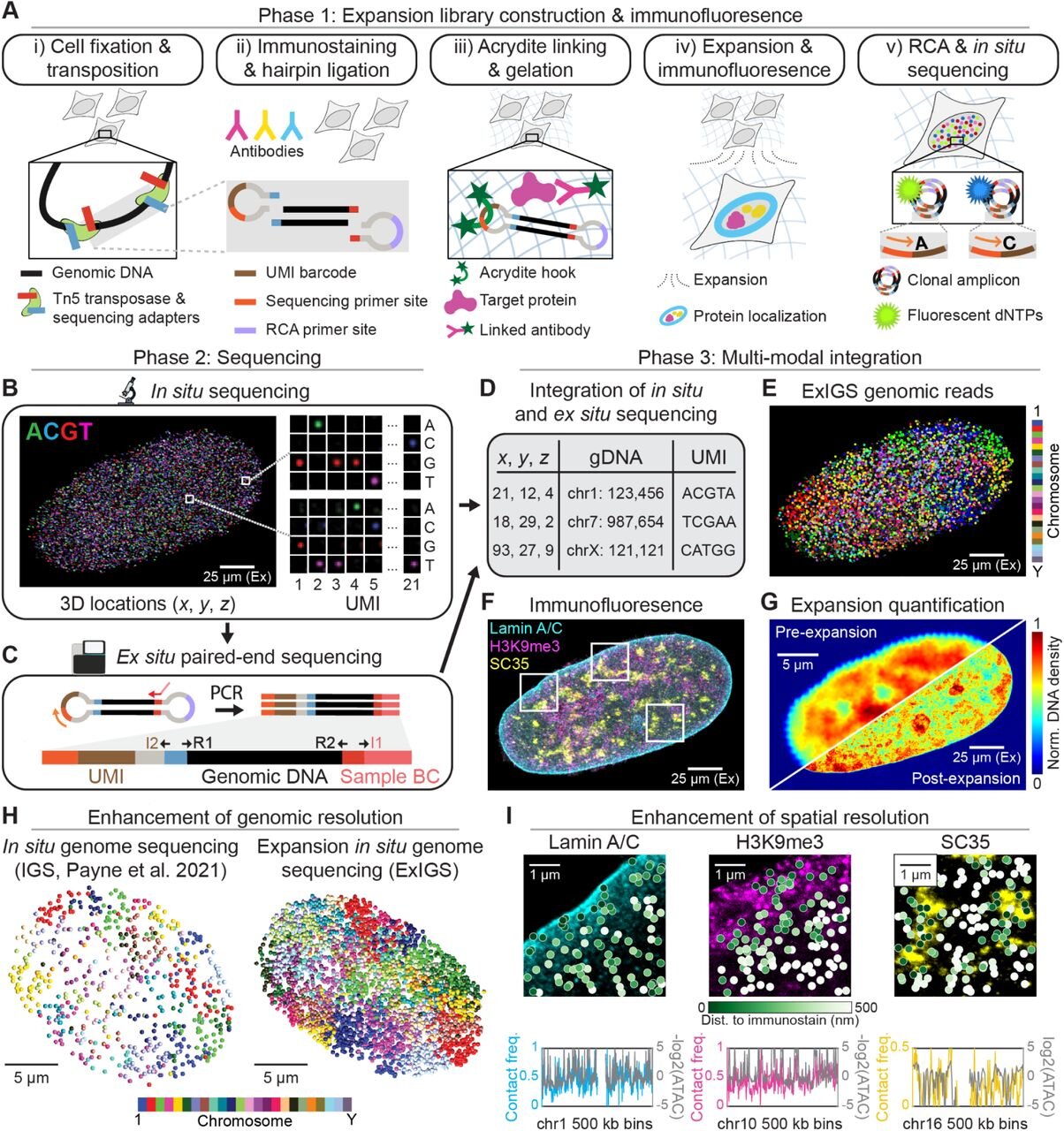
In ExIGS, expansion microscopy is integrated with in situ genome sequencing to simultaneously sequence genomic DNA and image nuclear proteins at nanoscale resolution within single cells.
The process begins with embedding fixed cells in a polyacrylate-based gel, which serves as a scaffold for expansion. Genomic DNA and proteins are chemically linked to the gel using DNA oligo hooks that bind to DNA hairpins attached to genomic fragments and proteins. These hooks contain chemical groups that form covalent bonds with the gel during polymerization.
Once embedded, the gel polymerizes around the fixed cellular components, immobilizing the DNA and proteins. Water is added to the gel causing it to swell uniformly by approximately 4.5 to 5.5 times in all directions. This expansion physically enlarges the cell while preserving the spatial relationships between molecules. To maintain the integrity of the newly expanded structure, the sample is then re-embedded in a secondary non-expandable acrylamide gel.
The expanded sample undergoes superresolution immunofluorescence imaging, allowing detailed visualization of nuclear proteins. Rolling circle amplification is then performed to generate clonal DNA amplicons, which are sequenced in situ. This amplification ensures accurate sequencing and localization of genomic DNA within the expanded nucleus.
The researchers detail testing of their innovation in a preprint paper, “Expansion in situ genome sequencing links nuclear abnormalities to hotspots of aberrant euchromatin repression,” on the bioRxiv preprint server.
By applying ExIGS to fibroblast cells from a patient with Hutchinson-Gilford progeria syndrome, researchers identified irregularities in nuclear lamins (proteins that give structure to the nucleus) that create hotspots of abnormal repression in active genomic regions, potentially compromising cell identity.
The study demonstrated that ExIGS significantly increases the number of genomic reads per nucleus and preserves 3D genome structure comparable to Hi-C sequencing data.
The technique provides a powerful new platform for exploring DNA-protein interactions, allowing researchers an unprecedented view of the molecular mechanisms. It could significantly enhance our understanding of a broad spectrum of diseases and genetic disorders, reveal the hidden interactions of cellular senescence in aging, and become a disruptive technology in biotechnology research labs.
More information:
Ajay S. Labade et al, Expansionin situgenome sequencing links nuclear abnormalities to hotspots of aberrant euchromatin repression, bioRxiv (2024). DOI: 10.1101/2024.09.24.614614
© 2024 Science X Network
Citation:
Expansion in situ genome sequencing innovation makes hidden DNA-protein interactions visible (2024, October 14)
retrieved 14 October 2024
from https://phys.org/news/2024-10-expansion-situ-genome-sequencing-hidden.html
This document is subject to copyright. Apart from any fair dealing for the purpose of private study or research, no
part may be reproduced without the written permission. The content is provided for information purposes only.