. DOI: 10.1038/s41467-024-48351-9")
Scintillators are detectors that make high-energy X-rays or particles visible through flashes of light to form an image. Their many applications include particle physics, medical imaging, X-ray security and more.
Despite their usefulness, however, scintillators have presented researchers with a conundrum. Until recently, scientists had to decide whether fast imaging or optimal performance was more important when choosing the appropriate scintillator technology for a particular experiment.
Scientists at the U.S. Department of Energy’s (DOE) Argonne National Laboratory may have found a way to resolve this dilemma. It involves a scintillator material composed of spherical particles that are 20 billionths of a meter in size. The research appears in Nature Communications.
Even though they are incredibly small, these nanoparticles have an intricate structure composed of a ball-like core of cadmium sulfide surrounded by a thin shell of cadmium selenide and a thicker shell of cadmium sulfide. Collaborating on this project were scientists from DOE’s Oak Ridge National Laboratory, Bowling Green State University (BGSU) and Northwestern University.
Due to quantum mechanical effects, these nanoparticles have valuable optical and electronic properties not possible with larger particles. The BGSU scientists synthesized these nanoparticles, called quantum shells, to form a close-knit lattice that constituted the scintillator material.
It is applicable to ultrafast radiation detection as well as the high-resolution imaging possible with X-ray light sources, such as the Advanced Photon Source (APS) at Argonne, a DOE Office of Science user facility.
An everyday application for scintillator technology can be found in a dentist’s office, where X-ray beams are shone through a patient’s mouth and onto a film of a reactive material that imprints an image of the teeth for the dentist to check for potential defects.
Although this kind of imaging is useful for dentists or doctors doing chest X-rays, it is a far cry from the power and precision needed for the nanoscale imaging such as that performed at the APS. That requires scintillator materials that are efficient, quick to respond, have great spatial resolution, are durable, and can be scaled to large sizes.
The research team’s recently developed quantum shells meet those criteria. “Quantum shells may be suitable for imaging in the dentist’s office, but they are much more well-suited for scintillators at a light source like the APS or for X-ray imaging of engines while they are running with liquids inside,” said Burak Guzelturk, a physicist in Argonne’s X-Ray Science Division.
“When traditional scintillators are excited by X-ray beams, they will emit light, and it will have some characteristic lifespan,” said Benjamin Diroll, a scientist in the Center for Nanoscale Materials, a DOE Office of Science user facility at Argonne.
“In some of them, it might be hundreds of nanoseconds, or it might be microseconds. The quantum shell scintillator achieves a single-digit nanosecond lifetime while preserving efficiency levels equal to traditional scintillators.”
Guzelturk compared quantum shells with another similar light-emitting material, quantum dots. “In a quantum dot, the light emission typically comes from the center part of the nano-object, and the color of light emitted depends on its size. On the other hand, in the quantum shells, the light emission does not originate from the core, but it’s actually the adjacent shell in the nanoparticle.”
The thickness of that shell determines how light is emitted. Scintillator material produced from quantum shells can deliver quick, well-defined imaging and long-term durability.
Classical scintillators tend to be quite thick. As a result, they can light up at the front or back or in the middle, which tends to blur the desired image. Quantum shell scintillators avoid that problem because they can be made as a thin film on a substrate material.
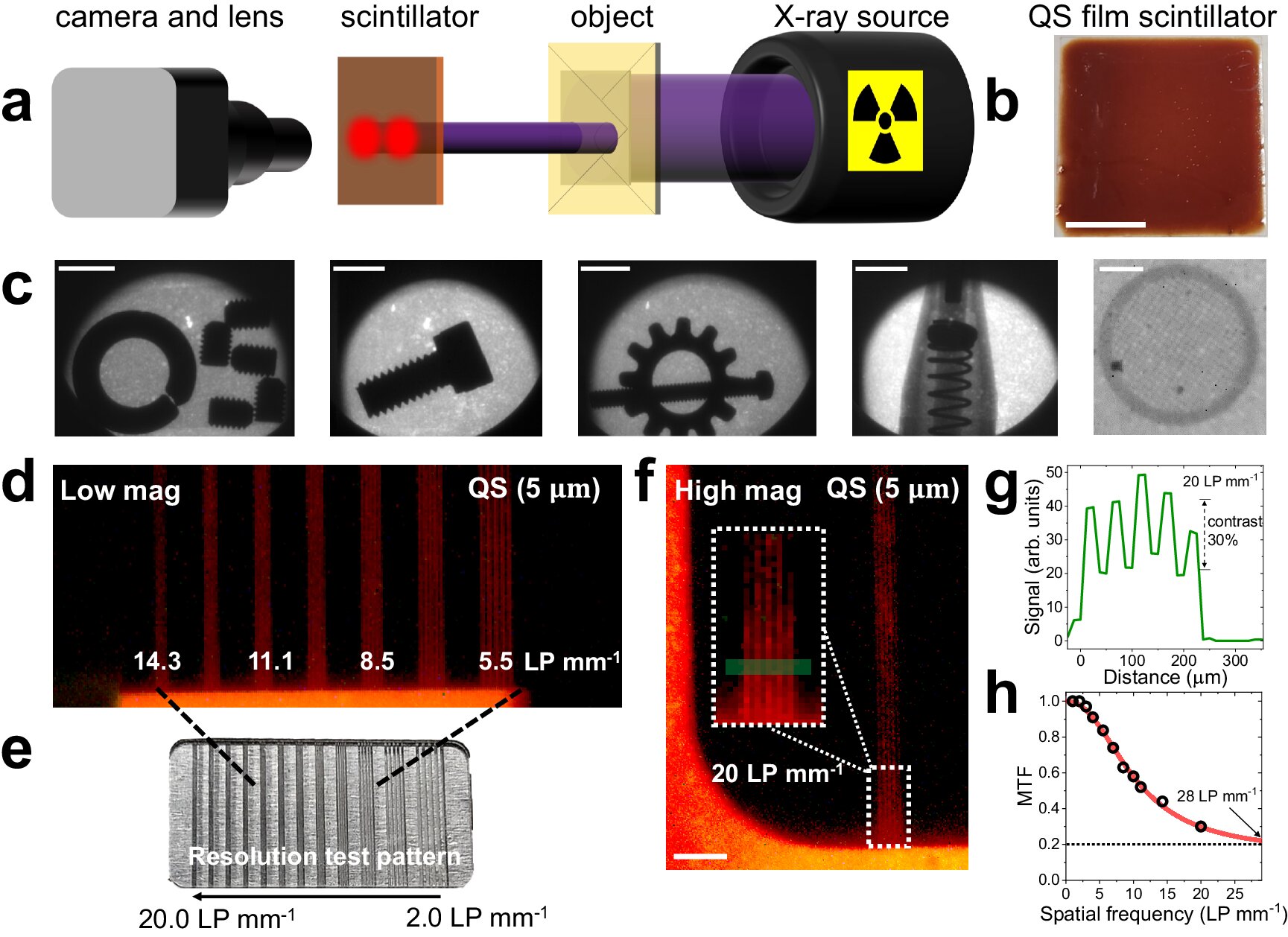
“Commercial scintillators that are made of lighter elements need to be millimeters thick,” explained Guzelturk. “In our case, we realized that we could make quantum shell scintillators much thinner, just a couple of micrometers, while achieving both strong X-ray absorption and high spatial resolution imaging.”
With the advent of quantum shell scintillators for high-resolution and ultrafast imaging, scientists are able to bypass the limitations of traditional scintillator technology. This pioneering work showcases the remarkable potential of these nanoscale quantum materials. By leveraging their unique optical and electronic properties, researchers can open new frontiers in fields ranging from particle physics to medical diagnostics.
More information:
Burak Guzelturk et al, Bright and durable scintillation from colloidal quantum shells, Nature Communications (2024). DOI: 10.1038/s41467-024-48351-9
Provided by
Argonne National Laboratory
Citation:
A quantum material could be the future of high-energy X-ray imaging and particle detection (2024, October 11)
retrieved 12 October 2024
from https://phys.org/news/2024-10-quantum-material-future-high-energy.html
This document is subject to copyright. Apart from any fair dealing for the purpose of private study or research, no
part may be reproduced without the written permission. The content is provided for information purposes only.