Why? Because it’s confounding to cure what we don’t understand, and the human brain, with its millions of neurons connected by a hundred trillion synapses, is almost hopelessly complex.
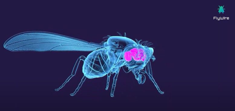
A Princeton-led team of neuroscientists has now made a massive step toward understanding the human brain, by building a neuron-by-neuron and synapse-by-synapse roadmap — scientifically speaking, a “connectome” — through the brain of an adult fruit fly (Drosophila melanogaster).
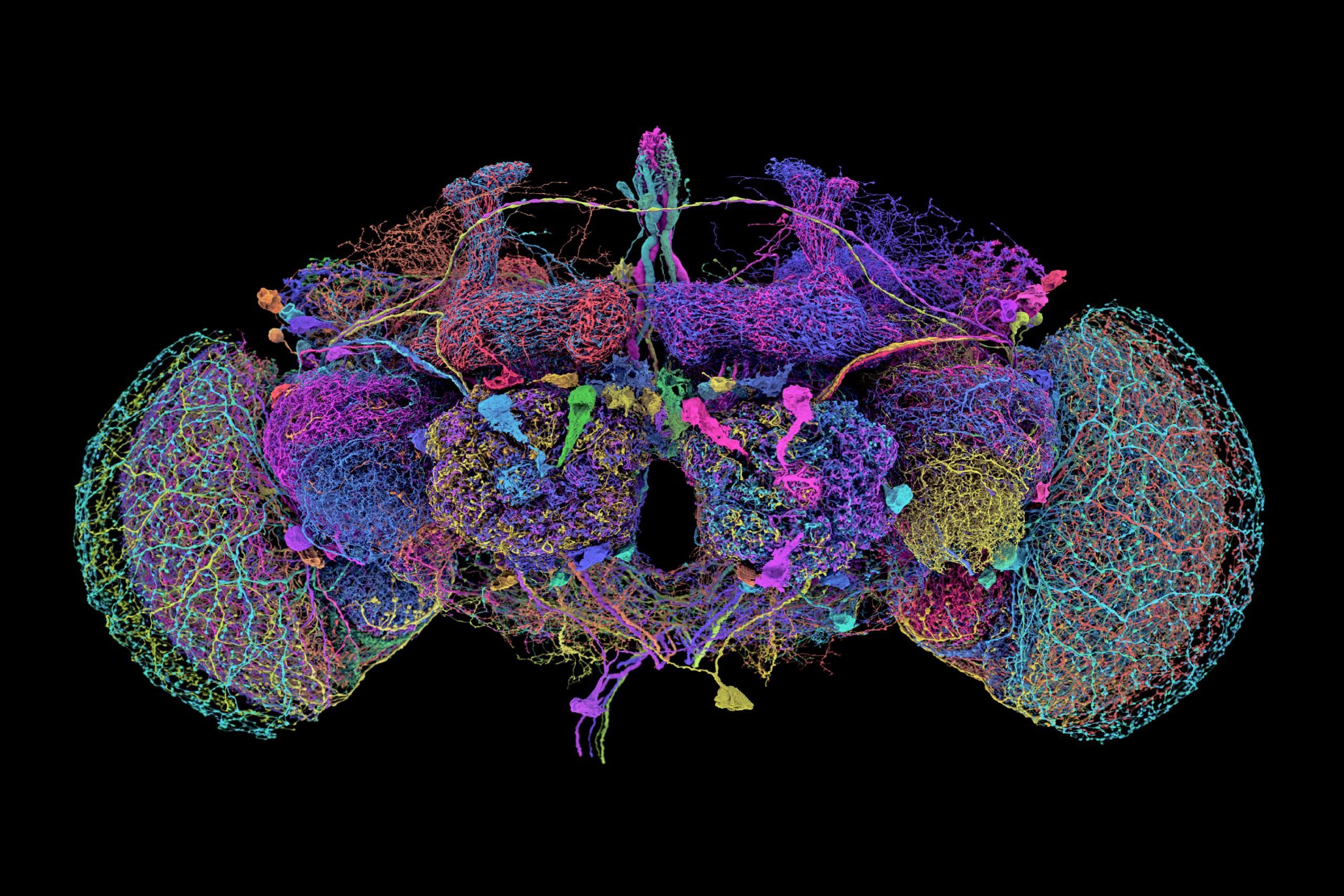
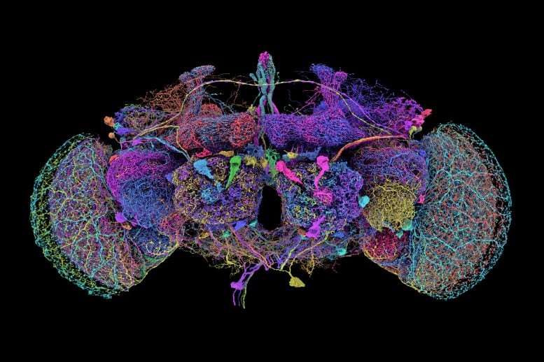
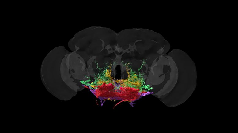
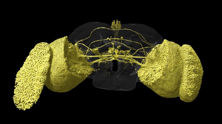
Credit: Amy Sterling / FlyWire / Princeton University
FlyWire Consortium built a complete connectome of every neuron in a Drosophila brain.
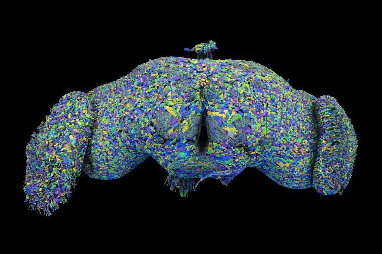
A Princeton-led team of scientists has created the first detailed connectome of an adult fruit fly brain, a complex network with almost 140,000 neurons. This significant step in neuroscience was featured in Nature and involved contributions from various global institutions, highlighting both the complexity of the fly’s brain and the potential insights it offers into human neurological diseases.
Groundbreaking Brain Mapping: A Connectome for the Adult Fruit Fly
Researchers led by Princeton University have constructed the first detailed neuron-by-neuron and synapse-by-synapse roadmap through the brain of an adult fruit fly (Drosophila melanogaster), achieving a major milestone in brain research. This study is the flagship article in the October 2 special issue of Nature, which is devoted to the new fruit fly “connectome.”
Previous efforts mapped the brain of a C. elegans worm, with its 302 neurons, and the brain of a larval fruit fly, which comprises about 3,000 neurons. However, the adult fruit fly is several orders of magnitude more complex, with nearly 140,000 neurons and approximately 50 million synapses connecting them.
Fruit flies share 60% of human DNA, and three in four human genetic diseases have a parallel in fruit flies. Understanding the brains of fruit flies is a stepping stone to understanding brains of larger more complex species, like humans.
Collaborative Effort in Neuroscience Research
“This is a major achievement,” said Mala Murthy, director of the Princeton Neuroscience Institute and, with Sebastian Seung, co-leader of the research team. “There is no other full brain connectome for an adult animal of this complexity.” Murthy is also Princeton’s Karol and Marnie Marcin ‘96 Professor of Neuroscience.
Princeton’s Seung and Murthy are co-senior authors on the flagship paper of the Nature issue, which includes a suite of nine related papers with overlapping sets of authors, led by researchers from Princeton University, the University of Vermont, the University of Cambridge, the University of California-Berkeley, UC-Santa Barbara, Freie Universität-Berlin, and the Max Planck Florida Institute for Neuroscience. The work was funded in part by the NIH’s BRAIN Initiative, the Princeton Neuroscience Institute’s Bezos Center for Neural Circuit Dynamics and McDonnell Center for Systems Neuroscience, and other public and private neuroscience institutes and funds, listed at the end of this document.
Building the Brain Atlas: The FlyWire Consortium
The map was developed by the FlyWire Consortium, which is based at Princeton University and made up of teams in more than 76 laboratories with 287 researchers around the world as well as volunteer gamers.
Sven Dorkenwald, the lead author on the flagship Nature paper, spearheaded the FlyWire Consortium.
“What we built is, in many ways, an atlas,” said Dorkenwald, a 2023 Ph.D. graduate of Princeton now at the University of Washington and the Allen Institute for Brain Science. “Just like you wouldn’t want to drive to a new place without Google Maps, you don’t want to explore the brain without a map. What we have done is build an atlas of the brain, and added annotations for all the businesses, the buildings, the street names. With this, researchers are now equipped to thoughtfully navigate the brain as we try to understand it.”
And just like a map that traces out every tiny alley as well as every superhighway, the fly connectome shows connections within the fruit fly brain at every scale.
Advances in AI and Neuroscience
The map was built from 21 million images taken of a female fruit fly brain by a team of scientists led by Davi Bock, then at the Howard Hughes Medical Institute’s Janelia Research Campus and now at the University of Vermont. Using an AI model built by researchers and software engineers working with Princeton’s Sebastian Seung, the lumps and blobs in those images were turned into a labeled, three-dimensional map. Instead of keeping their data confidential, the researchers opened their in-progress neural map to the scientific community from the beginning.
“Mapping the whole brain has been made possible by advances in AI computing. It would have not been possible to reconstruct the entire wiring diagram manually. This is a display of how AI can move neuroscience forward,’ said Prof. Sebastian Seung, one of the co-leaders of the research and Princeton’s Evnin Professor in Neuroscience and a professor of computer science.
“Now that we have this brain map, we can close the loop on which neurons relate to which behaviors,” said Dorkenwald.
The development could lead to tailored treatments to brain diseases.
“In many respects, it (the brain) is more powerful than any human-made computer, yet for the most part we still do not understand its underlying logic,” said John Ngai, director of the U.S. National Institutes of Health’s BRAIN Initiative, which provided partial funding for the FlyWire project. “Without a detailed understanding of how neurons connect with one another, we won’t have a basic understanding of what goes right in a healthy brain or what goes wrong in disease.”
For more on this breakthrough:
Reference: “Neuronal wiring diagram of an adult brain” by Sven Dorkenwald, Arie Matsliah, Amy R. Sterling, Philipp Schlegel, Szi-chieh Yu, Claire E. McKellar, Albert Lin, Marta Costa, Katharina Eichler, Yijie Yin, Will Silversmith, Casey Schneider-Mizell, Chris S. Jordan, Derrick Brittain, Akhilesh Halageri, Kai Kuehner, Oluwaseun Ogedengbe, Ryan Morey, Jay Gager, Krzysztof Kruk, Eric Perlman, Runzhe Yang, David Deutsch, Doug Bland, Marissa Sorek, Ran Lu, Thomas Macrina, Kisuk Lee, J. Alexander Bae, Shang Mu, Barak Nehoran, Eric Mitchell, Sergiy Popovych, Jingpeng Wu, Zhen Jia, Manuel A. Castro, Nico Kemnitz, Dodam Ih, Alexander Shakeel Bates, Nils Eckstein, Jan Funke, Forrest Collman, Davi D. Bock, Gregory S. X. E. Jefferis, H. Sebastian Seung, Mala Murthy and The FlyWire Consortium, 2 October 2024, Nature.
DOI: 10.1038/s41586-024-07558-y
This research was supported by the National Institutes of Health (NIH) BRAIN Initiative (RF1 MH117815, RF1 MH129268, 1RF1MH120679-01 and U24 NS126935) and National Institute Of Neurological Disorders And Stroke (NINDS) (RF1NS121911); the Princeton Neuroscience Institute’s Bezos Center for Neural Circuit Dynamics and McDonnell Center for Systems Neuroscience; Google; the Allen Institute for Brain Science; the National Science Foundation (NSF Neuronex2 2014862, Neuronex2 MRC MC_EX_MR/T046279/1, MRC MC-U105188491, PHY-1734030); Wellcome Trust Collaborative Award (203261/Z/16/Z and 220343/Z/20/Z); a Marie Skłodowska-Curie postdoctoral fellowship (H2020-WF-01-2018-867459); the Portuguese Research Council (Grant PTDC/MED-NEU/4001/2021); and the Intelligence Advanced Research Projects Activity (IARPA) via the Department of Interior (DOI) (D16PC0005).